Although Aspergillus oryzae belongs to the lower eukaryotic family of microorganisms just as the yeasts, it is a multicellular organism and grows in a filamentous form with many compartments in a row unlike the yeast being an egg-shaped single cell. Due to this unique form, the number and the structures of the organelles at the tip cell of hyphae are greatly different from those of the basal cells. Moreover, it is thought that the organelles, such as the nuclei and the vacuoles, are arranged in suitable positions in conidium cells which is important for tane-koji to maintain the state where they can bud off under suitable conditions. While a lot of research is focused on the enzyme production and growth of Aspergillus oryzae, there has been almost no research on the structural analysis of its cellular organelles. We think that it is important to understand the biology of Aspergillus oryzae cells both for fundamental purpose and for its application in brewing and enzyme production. Therefore research on visualizing the organelles and their dynamics in Aspergillus oryzae forms an important part of our study. Fig. 1 typically shows the organelles in Aspergillus oryzae cell, based on the available knowledge on their structure in filamentous fungal cell.

ER:Endoplasmic reticulum G:Golgi body M:Mitochondria N:nucleus V:vacuole
Fig.1
In filamentous fungi, the nuclei are located everywhere from hyphae to conidia, and their accurate localization is necessary for hyphal growth and conidium formation. Moreover, it is surmised that nuclear transport is greatly involved in Aspergillus oryzae activities in brewing or industrial use. We visualized the nuclei in Aspergillus oryzae by expressing the Histone H2B and EGFP (Enhanced Green Fluorescent Protein) fusion protein (Fig. 2).
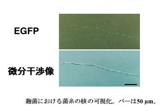
the upper ; EGFP.
the lower ;differential interference image.
The length of the bar is 50 μm
Fig. 2 Visualization of nuclei in Aspergillus oryzae hypha
It is possible to observe nuclei within a living cell by using EGFP, unlike DAPI dyeing used so far for nuclear visualization. By observing the tip cells of hyphae sequentially, we found that the shape of many nuclei was a little long and slender, and we obtained a dynamic image that the nuclear movement was irregular in the direction of the tip, or in the direction of the base, and finally it was accompanied by hyphal growth. (Fig.3)
Moreover, we also succeeded in observing mitosis sequentially. (Fig.4)
Aspergillus oryzae forms the asexual spore called a conidium like the other filamentous fungi, but it is multinucleate. (Fig.5) Since, it is essential that Aspergillus oryzae has hereditary stability in the long history of brewing, it is thought that Aspergillus oryzae which has multinucleate conidia has been selected for quality maintenance of koji over many years. FACS (Fluorescence-Activated Cell Sorter) is the system which irradiates each cell with the laser beam, detects dispersion light and fluorescence, and measures it as numerical values. Our laboratory introduced FACS into research on filamentous fungi for the first time, and used it for the analysis of the nuclear number in conidia. We have been trying to mutate conidia whose nuclei were visualized by EGFP and screen mutant strains which formed uninucleate conidia a lot by FACS, and we have succeeded in acquiring the mutant strains whose 80% or more conidia were uninucleate. It is expected this research not only provides information on multinucleate conidium formation mechanism in Aspergillus oryzae but also helps in acquiring homokaryotic strains.
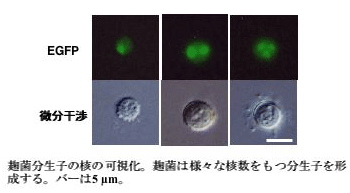
Visualization of nuclei in Aspergillus oryzae conidia.
Aspergillus oryzae forms conidia with varied nuclear numbers.
The length of the bar is 5μm
Fig.5